The mechanisms of cancer are discussed by analyzing the characteristics of the functional state and biological behavior of the abnormal nuclear cells. The abnormal nuclear cells with abnormal nuclear structure and function are a kind of sick cell or functional defect cells having existed in human body for a long time. The abnormal nuclear cells are resulted from the nuclear damage caused by the radiation, viruses and various carcinogenic compounds. Some of genes in human body are expressed, some are not expressed for life. The expressional genes are functional genes, the genes never expressed for life in human body are dormant genes or sealed genes. The nuclear damages destroy cell state of differentiation, affect gene expressional regulation and change gene expressional profiling, resulting in loss of expression of the functional genes and reactivation of the sealed genes; which finally leads to cancer, aging and other chronic refractory diseases. The cancer is not resulted from the genetic mutations or chromosomal aberrations, but rather the reactivation of genes involved in proliferation due to the nuclear damage. The biological characteristics of the cancer cells, such as the shedding and metastasis, immune tolerance, uncontrolled, loss of contact inhibition function and so on, all originate from the nuclear aberrant cells. The nuclear damage can trigger the genes that drive mitosis, leading to cancer. Thus, re-sealing the several genes that trigger the proliferation may completely prevent or cure cancers.
| Published in | Cancer Research Journal (Volume 13, Issue 2) |
| DOI | 10.11648/j.crj.20251302.15 |
| Page(s) | 71-80 |
| Creative Commons |
This is an Open Access article, distributed under the terms of the Creative Commons Attribution 4.0 International License (http://creativecommons.org/licenses/by/4.0/), which permits unrestricted use, distribution and reproduction in any medium or format, provided the original work is properly cited. |
| Copyright |
Copyright © The Author(s), 2025. Published by Science Publishing Group |
Abnormal Nuclear Cell, Abnormal Nuclear, Nuclear Damage, Cancer, Gene, Regulation of Gene Expression, Functional Gene, Sealed Gene
Items | Normal cells | Abnormal nuclear cells |
|---|---|---|
Morphology and structure of nucleus | normal | abnormal |
Nuclear function (replication, transcription, ribosome synthesis) | normal | abnormal |
Gene mutation | no or yes | yes |
Chromosome aberration | no | yes |
Gene expression regulation | normal | abnormal |
Cell differentiation state | normal | unstable or disrupted |
Functional genes | normal expression | abnormal expression |
Seal genes (dormant gene) | not expressed (in a sealed state) | start to express |
DNA transcription-protein synthesis function | normal | abnormal |
RNA | Ribonucleic Acid |
DNA | Deoxyribonucleic Acid |
ROS | Reactive Oxygen Species |
ANC | Abnormal Nuclear Cells |
SVG | Survival Genes |
FG | Functional Genes |
DG | Dormant Genes |
HG | Helper Genes |
SG | Sealed Genes |
FR | Free Radicals |
RG | Role Genes |
| [1] | Xia C, Dong X, Li H, et al. Cancer statistics in China and United States, 2022: profiles, trends, and determinants [J]. Chin Med J (Engl). 2022, 135(5): 584-590. |
| [2] | Siegel RL, Giaquinto AN, Jemal A. Cancer statistics, 2024 [J]. CA Cancer J Clin. 2024, 74(1): 12-49. |
| [3] | Wu Y, He S, Cao M, et al. Comparative analysis of cancer statistics in China and the United States in 2024. Chin Med J (Engl). 2024, 137(24): 3093-3100. |
| [4] | Basu AK. DNA Damage, Mutagenesis and Cancer [J]. Int J Mol Sci, 2018, 19(4): 970. |
| [5] | Graham TA, Sottoriva A. Measuring cancer evolution from the genome [J]. J Pathol, 2017, 241(2): 183-191. |
| [6] | Xueqian Yan, Li Liu, Yueru Ji, et al. Relationship between serum miR-155-5p and miR-146b-5p and chromosome aberration and prognosis of secondary acute myeloid leukemia [J]. Medical Journal of the Chinese People's Armed Police Force. 2024, 35(01): 14-20+24. |
| [7] | Qiongyu Li. The study of ribosome biosynthesis involved in ribosomal proteins in cancer [J]. Chinese Journal of Modern Drug Application, 2024, 18(04): 174-180. |
| [8] | Jiramongkol Y, Lam EW. FOXO transcription factor family in cancer and metastasis [J]. Cancer Metastasis Rev. 2020, 39(3): 681-709. |
| [9] | Geden MJ, Romero SE, Deshmukh M. p53 is required for nuclear but not mitochondrial DNA damage-induced degeneration [J]. CellDeath Dis, 2021, 12(1): 104. |
| [10] | Abdel Rhman M, Pmo O. Potential therapeutic applications of microRNAs in cancer diagnosis and treatment: Sharpening adouble-edged sword? [J]. Eur J Pharmacol, 2022, 932: 175210. |
| [11] | Jie Jiang, Zai Luo, Haoliang Zhang, et al. Circular RNA-Encoded Proteins in Gastrointestinal Cancer [J]. Acta Academiae Medicinae Sinicae, 2024, 46(01): 72-81. |
| [12] | Judasz E, Lisiak N, Kopczyński P, et al. The role of telomerase in breast cancer's response to therapy [J]. Int J Mol Sci, 2022, 23(21): 12844. |
| [13] | Engin AB, Engin A. The connection between cell fate and telomere [J]. Adv Exp Med Biol, 2021, 1275: 71-100. |
| [14] | Taylor AMR, Rothblum-Qviatt C, Ellis NA, et al. Coromosome instability syndromes [J]. Nat Rev Dis Primers, 2019, 5(1): 64. |
| [15] | Yunxiao Xie, Na Ding. Analysis of the Effect of Methylation Alterations on Enhancer Activity in Cancer [J]. International Journal of Geriatrics, 2024, 45(02): 193-198. |
| [16] | Saghafinia S, Mina M, Riggi N, et al. Pan-cancer landscape of aberrant DNA methylation across human tumors [J]. Cell Rep, 2018, 25(4): 1066-1080.e8. |
| [17] | Zappe K, Cichna-Markl M. Aberrant DNA methylation of ABC transporters in cancer [J]. Cells, 2020, 9(10): 2281. |
| [18] | Zafon C, Gil J, Pérez-González B, et al. DNA methylation inthyroid cancer [J]. Endocr Relat Cancer, 2019, 26(7): R415-R439. |
| [19] | Capuozzo M, Santorsola M, Bocchetti M, et al. p53: From Fundamental Biology to Clinical Applications in Cancer [J]. Biology (Basel). 2022, 11(9): 1325. |
| [20] | Martinez-Fundichely A, Dixon A, Khurana E. Modeling tissue-specific breakpoint proximity of structural variations from whole-genomes to identify cancer drivers [J]. Nat Commun. 2022, 13(1): 5640. |
| [21] | Tiwari B, Jones AE, Caillet CJ, et al. p53 directly represses human LINE1 transposons [J]. Genes Dev. 2020, 34(21-22): 1439-1451. |
| [22] | Jingjing Guo, Di Mou, Wenwen Yu, et al. Study on the Biological Function of Abemecilib in Inhibiting the Proliferation, Invasion and Migration of Small Cell Lung Cancer with High c-Myc Expression [J]. Chinese Journal of Lung Cancer, 2023, 26(02): 105-112. |
| [23] | Maojin Li, Anyi Hu. The research progress and biological significance of abnormality of cell nucleus [J]. China Occupational Medicine, 2021, 48(6): 708-711. |
| [24] | Ashley DJ. The two "hit" and multiple "hit" theories ofcarcinogenesis [J]. Br J Cancer, 1969, 23(2): 313-328. |
| [25] | Jabbour E, Short NJ, Jain N, et al. The evolution of acute lymphoblastic leukemia research and therapy at MD Anderson over four decades [J]. J Hematol Oncol. 2023, 16(1): 22. |
| [26] | Saleh K, Fernandez A, Pasquier F. Treatment of Philadelphia Chromosome-Positive Acute Lymphoblastic Leukemia in Adults [J]. Cancers (Basel). 2022, 14(7): 1805. |
| [27] | Qiang Lu, Qiuling Zhang, Xiujuan Hong. Relationships between chromosome aberration, blood routine, and cumulative dose in 718 radiation workers [J]. Industrial Health and Occupational Diseases, 2023, 49(06): 497-500. |
| [28] | Rageul J, Kim H. Fanconi anemia and the underlying causes of genomic instability [J]. Environ Mol Mutagen. 2020, 61(7): 693-708. |
| [29] | García-de-Teresa B, Rodríguez A, Frias S. Chromosome Instability in Fanconi Anemia: From Breaks to Phenotypic Consequences [J]. Genes (Basel). 2020, 11(12): 1528. |
| [30] | Hongling Ou, Wenchao Ai, Yan Wang, et al. Advances in research and application of ionizing radiation biomarkers [J]. Chinese Journal of Pharmacology and Toxicology, 2024, 38(01): 70-78. |
| [31] | Devadoss S, Raveendranath MC, Kathiresan TS, et al. Genotoxic effect of various forms of tobacco on oral buccal mucosa and nuclear changes as a biomarker [J]. J Pharm Bio allied Sci, 2021, 13(Suppl 2): S1141-S1148. |
| [32] | Druzhinin V, Bakanova M, Fucic A, et al. Lymphocytes with multiple chromosomal damages in a large cohort of West Siberia residents: results of long-term monitoring [J]. Mutat Res, 2016, 784-785: 1-7. |
| [33] | Lei Wang, Xiaomin Xu, Yanhui Zhang, et al. Effects of Astragalus Polysaccharide on X-ray Radiation-induced Nucleus, Chromosome and DNA Damage in Bone Mesenchymal Stem Cells [J]. China Cancer, 2018, 27(9): 708-714. |
| [34] | Nembhard WN, McElfish PA, Ayers B, et al. Nuclear radiation and prevalence of structural birth defects among infants born to women from the Marshall Islands [J]. Birth Defects Res, 2019, 111(16): 1192-1204. |
| [35] | Bouza E, Martín Jiménez M, Alemany L, et al. Overview of virus and cancer relationships [J]. Position paper. Rev Esp Quimioter. 2021, 34(6): 525-555. |
| [36] | Zhang L, Richards A, Barrasa MI, et al. Reverse-transcribedSARS-CoV-2 RNA can integrate into the genome of culturedhuman cells and can be expressed in patient-derived tissues [J]. Proc Natl Acad Sci U S A, 2021, 118(21): e2105968118. |
| [37] | Rodriguez-Muñoz M, Anglada T, Genescà A. A matter of wrapper: Defects in the nuclear envelope of lagging and bridging chromatin threatens genome integrity. Semin Cell Dev Biol. 2022, 123: 124-130. |
| [38] | Maojin Li, Rugang Wang, Anyi Hu. The analysis of lymphocytes with nuclei abnormalities in peripheral blood of patients with cancers [J]. Journal of Public Health and Preventive Medicine, 2019, 30(1): 27-31. |
| [39] | Caracausi M, Piovesan A, Antonaros F, et al. Systematic identification of human housekeeping genes possibly useful as references ingene expression studies [J]. Mol Med Rep, 2017, 16(3): 2397-2410. |
| [40] | Joshi CJ, Ke W, Drangowska-Way A, et al. What are housekeeping genes? [J]. PLoS Comput Biol, 2022, 18(7): e1010295. |
| [41] | Saini J, Sharma PK. Clinical, prognostic and therapeutic significance of heat shock proteins in cancer [J]. Curr Drug Targets, 2018, 19(13): 1478-1490. |
| [42] | De Jong TV, Moshkin YM, Guryev V. Gene expression variability: the other dimension in transcriptome analysis [J]. Physiol Genomics, 2019, 51(5): 145-158. |
| [43] | Signor SA, Nuzhdin SV. The evolution of gene expression in cisand trans [J]. Trends Genet, 2018, 34(7): 532-544. |
| [44] | Manda NK, Golla U, Sesham K, et al. Tuning between nuclearorganization and functionality in health and disease [J]. Cells, 2023, 12(5): 706. |
| [45] | Funk L, Su KC, Ly J, et al. The phenotypic landscape of essential human genes [J]. Cell, 2022, 185(24): 4634-4653.e22. |
| [46] | Bonora G, Ramani V, Singh R, et al. Single-cell landscape of nuclear configuration and gene expression during stem cell differentiation and X inactivation [J]. Genome Biol. 2021, 22(1): 279. |
| [47] | Ng HL, Quail E, Cruickshank MN, et al. To be, or notch to be: mediating cell fate from embryogenesis to lymphopoiesis [J]. Biomolecules, 2021, 11(6): 849. |
| [48] | Jaiswal SK, Raj S, DePamphilis ML. Developmental Acquisition of p53 Functions [J]. Genes (Basel). 2021, 12(11): 1675. |
| [49] | Gordeeva O. TGFβ Family Signaling Pathways in Pluripotent and Teratocarcinoma Stem Cells' Fate Decisions: Balancing Between Self-Renewal, Differentiation, and Cancer [J]. Cells. 2019, 8(12): 1500. |
| [50] | Kim YJ, Tamadon A, Kim YY, et al. Epigenetic Regulation of Cardiomyocyte Differentiation from Embryonic and Induced Pluripotent Stem Cells [J]. Int J Mol Sci. 2021, 22(16): 8599. |
| [51] | Wu Z, Guan KL. Hippo signaling in embryogenesis and development [J]. Trends Biochem Sci, 2021, 46(1): 51-63. |
| [52] | Leu S. The role and regulation of Pnn in proliferative and non-dividing cells: Form embryogenesis to pathogenesis [J]. Biochem Pharmacol, 2021, 192: 114672. |
| [53] | Sigvardsson M. Molecular regulation of differentiation in early B-lymphocyte development [J]. Int J Mol Sci, 2018, 19(7): 1928. |
| [54] | Shparberg RA, Glover HJ, Morris MB. Modeling Mammalian Commitment to the Neural Lineage Using Embryos and Embryonic Stem Cells [J]. Front Physiol. 2019, 10: 705. |
| [55] | Kovacs MT, Vallette M, Wiertsema P, et al. DNA damage induces nuclear envelope rupture through ATR-mediated phosphorylation of lamin A/C. Mol Cell. 2023, 83(20): 3659-3668.e10. |
| [56] | Bonora M, Missiroli S, Perrone M, et al. Mitochondrial Control of Genomic Instability in Cancer. Cancers (Basel). 2021, 13(8): 1914. |
| [57] | Janssen A, Breusegem SY, Larrieu D. Current methods and pipelines for image-based quantitation of nuclear shape and nuclear envelope bbnormalities [J]. Cells, 2022, 11(3): 347. |
| [58] | Abdalla F, Boder J, Markus R, et al. Correlation of nuclear morphometry of breast cancer in histological sections with clinic pathological features and prognosis [J]. Anticancer Res, 2009, 29(5): 1771-1776. |
| [59] | Bian Z, Wang L, Fan R, et al. Genetic predisposition, modifiable lifestyles, and their joint effects on human lifespan: evidence from multiple cohort studies. BMJ Evid Based Med. 2024, 29(4): 255-263. |
| [60] | Johnston RA, Aracena KA, Barreiro LB, et al. DNA methylation-environment interactions in the human genome. Elife. 2024 Feb 26; 12: RP89371. |
| [61] | Minqiang Xie. Research progress in the treatment of nasopharyngeal carcinoma [J]. Chinese Journal of Otorhinolaryngology-skull Base Surgery, 2023, 29(06): 1-10. |
| [62] | Cronin KA, Scott S, Firth AU, et al. Annual report to the nation on the status of cancer, part 1: National cancer statistics [J]. Cancer. 2022, 128(24): 4251-4284. |
| [63] | Yang X, Chen H, Sang S, et al. Burden of All Cancers Along With Attributable Risk Factors in China From 1990 to 2019: Comparison With Japan, European Union, and USA [J]. Front Public Health. 2022, 10: 862165. |
| [64] | Zafar A, Khan MJ, Abu J, et al. Revolutionizing cancer care strategies: immunotherapy, gene therapy, and molecular targeted therapy [J]. Mol Biol Rep. 2024, 51(1): 219. |
| [65] | Shoaib S, Islam N, Yusuf N. Phytocompounds from the Medicinal and Dietary Plants: Multi-target Agents for Cervical Cancer Prevention and Therapy [J]. Curr Med Chem. 2022, 29(26): 4481-4506. |
| [66] | Jiasi Yan. Research progress of biotherapy and biotechnology drugs [J]. Modern Chemical Research, 2023,(05): 8-10. |
| [67] | Brown DW, Beatty PH, Lewis JD. Molecular Targeting of the Most Functionally Complex Gene in Precision Oncology: p53 [J]. Cancers (Basel). 2022, 14(21): 5176. |
| [68] | Qiaomei Wu, Jinglin Yang, Ya Peng, et al. Analysis of occurrence and symptom groups of lung cancer patients dur-ing immunotherapy [J]. China Modern Medicine, 2024, 31(04): 144-148. |
| [69] | Fei Duan, Yi Chen, Guanjin Cha, Xunhuang Duan. Effects of miR-340-5p targeting regulation of ARID1A on activity, migration and invasion of thyroid cancer [J]. Chinese Journal of Gerontology, 2024, 44(06): 1480-1485. |
| [70] | Sammons MA, Nguyen TT, McDade SS, et al. Tumor suppressor p53: from engaging DNA to target gene regulation [J]. Nucleic Acids Res. 2020, 48(16): 8848-8869. |
APA Style
Li, M. (2025). Cancers Are Derived from the Disruption of Cell Differentiation. Cancer Research Journal, 13(2), 71-80. https://doi.org/10.11648/j.crj.20251302.15
ACS Style
Li, M. Cancers Are Derived from the Disruption of Cell Differentiation. Cancer Res. J. 2025, 13(2), 71-80. doi: 10.11648/j.crj.20251302.15
@article{10.11648/j.crj.20251302.15,
  author = {Maojin Li},
  title = {Cancers Are Derived from the Disruption of Cell Differentiation
},
  journal = {Cancer Research Journal},
  volume = {13},
  number = {2},
  pages = {71-80},
  doi = {10.11648/j.crj.20251302.15},
  url = {https://doi.org/10.11648/j.crj.20251302.15},
  eprint = {https://article.sciencepublishinggroup.com/pdf/10.11648.j.crj.20251302.15},
  abstract = {The mechanisms of cancer are discussed by analyzing the characteristics of the functional state and biological behavior of the abnormal nuclear cells. The abnormal nuclear cells with abnormal nuclear structure and function are a kind of sick cell or functional defect cells having existed in human body for a long time. The abnormal nuclear cells are resulted from the nuclear damage caused by the radiation, viruses and various carcinogenic compounds. Some of genes in human body are expressed, some are not expressed for life. The expressional genes are functional genes, the genes never expressed for life in human body are dormant genes or sealed genes. The nuclear damages destroy cell state of differentiation, affect gene expressional regulation and change gene expressional profiling, resulting in loss of expression of the functional genes and reactivation of the sealed genes; which finally leads to cancer, aging and other chronic refractory diseases. The cancer is not resulted from the genetic mutations or chromosomal aberrations, but rather the reactivation of genes involved in proliferation due to the nuclear damage. The biological characteristics of the cancer cells, such as the shedding and metastasis, immune tolerance, uncontrolled, loss of contact inhibition function and so on, all originate from the nuclear aberrant cells. The nuclear damage can trigger the genes that drive mitosis, leading to cancer. Thus, re-sealing the several genes that trigger the proliferation may completely prevent or cure cancers.
},
 year = {2025}
}
TY  - JOUR
T1  - Cancers Are Derived from the Disruption of Cell Differentiation

AU  - Maojin Li
Y1  - 2025/06/23
PY  - 2025
N1  - https://doi.org/10.11648/j.crj.20251302.15
DO  - 10.11648/j.crj.20251302.15
T2  - Cancer Research Journal
JF  - Cancer Research Journal
JO  - Cancer Research Journal
SP  - 71
EP  - 80
PB  - Science Publishing Group
SN  - 2330-8214
UR  - https://doi.org/10.11648/j.crj.20251302.15
AB  - The mechanisms of cancer are discussed by analyzing the characteristics of the functional state and biological behavior of the abnormal nuclear cells. The abnormal nuclear cells with abnormal nuclear structure and function are a kind of sick cell or functional defect cells having existed in human body for a long time. The abnormal nuclear cells are resulted from the nuclear damage caused by the radiation, viruses and various carcinogenic compounds. Some of genes in human body are expressed, some are not expressed for life. The expressional genes are functional genes, the genes never expressed for life in human body are dormant genes or sealed genes. The nuclear damages destroy cell state of differentiation, affect gene expressional regulation and change gene expressional profiling, resulting in loss of expression of the functional genes and reactivation of the sealed genes; which finally leads to cancer, aging and other chronic refractory diseases. The cancer is not resulted from the genetic mutations or chromosomal aberrations, but rather the reactivation of genes involved in proliferation due to the nuclear damage. The biological characteristics of the cancer cells, such as the shedding and metastasis, immune tolerance, uncontrolled, loss of contact inhibition function and so on, all originate from the nuclear aberrant cells. The nuclear damage can trigger the genes that drive mitosis, leading to cancer. Thus, re-sealing the several genes that trigger the proliferation may completely prevent or cure cancers.

VL  - 13
IS  - 2
ER  -
Department of Physical Examination, the Beijing Prevention and Treatment Hospital of Occupational Disease / Beijing Institute of Occupational Disease Prevention and Treatment, Beijing, China
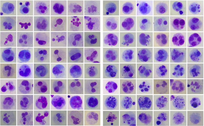
Figure 1. The abnormal nuclear cells.
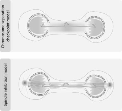
Figure 2. The model for the nuclear anomalies.
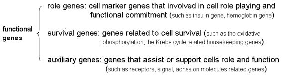
Figure 3. The functional genes.
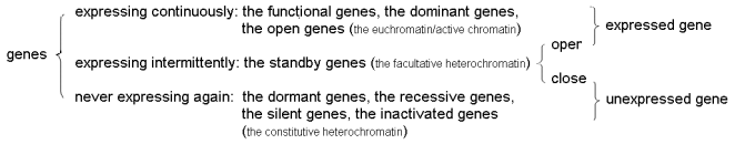
Figure 4. The gene expressional state.
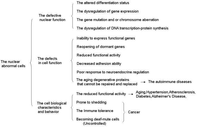
Figure 5. The cell biological characteristics and behavior of the abnormal nuclear cells.
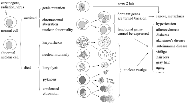
Figure 6. The chronic intractable diseases probably derived from the nuclear damage.